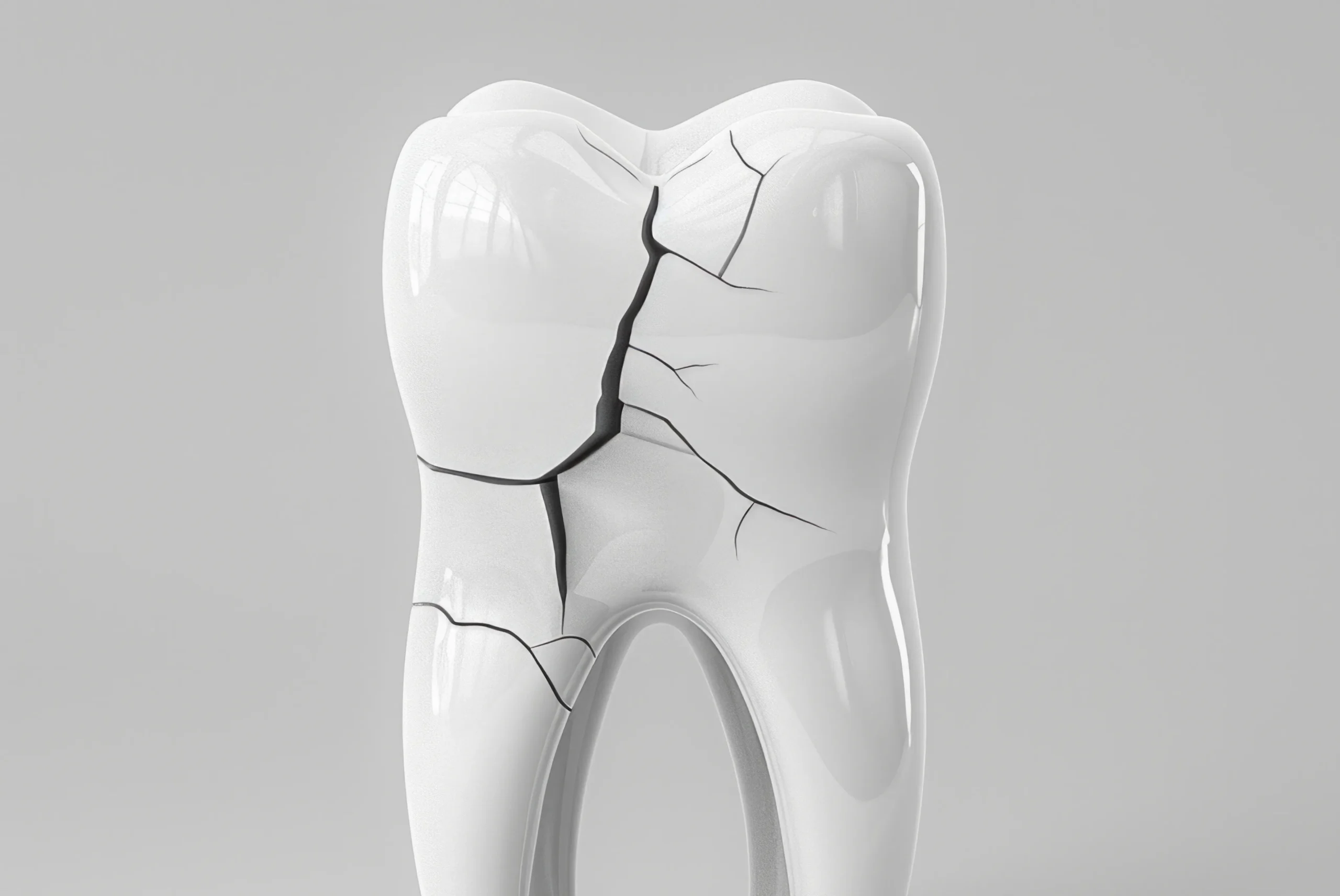
Hidden Cracks: The Silent Culprits Behind Your Masticatory Pain
Beneath the surface of seemingly healthy teeth lies a hidden issue that challenges both comfort and function. Often unnoticed until it disrupts daily activity, this condition emerges silently, bringing fleeting discomfort and increased sensitivity, ultimately posing a threat to dental health and requiring timely, targeted intervention.
Decoding the Signals of Intermittent Discomfort
The Enigma of the "Electric Shock" Sensation
The discomfort caused by a fractured tooth is notoriously elusive, often defying the standard descriptions of dental pain. Unlike the dull, constant throbbing associated with a deep cavity or an abscess, the symptoms of a structural crack are erratic and unpredictable. Patients frequently report a sharp, sudden sensation—likened to a jolt of electricity—that pierces through the jaw during a meal, only to vanish as quickly as it appeared. One day, biting into a piece of toast might trigger an agonizing spike of pain; the next day, chewing the same food on the same side might feel perfectly normal.
This unpredictability is the primary source of stress for many patients. The intermittent nature of the symptoms leads to a dangerous cycle of rationalization. It is common to think, "It hurt yesterday, but I feel fine today, so perhaps it has healed." Consequently, many people delay seeking professional help, assuming the problem was a temporary fluke. However, unlike skin or bone, dental structures do not possess the biological capacity to regenerate or knit themselves back together. A microscopic fissure within the enamel or dentin will not resolve on its own. Instead, it operates silently, often deepening with every meal, turning a minor structural flaw into a significant dental emergency. Understanding that this "come and go" pattern is a hallmark of structural compromise, rather than a sign of healing, is crucial for saving the tooth.
Why Letting Go Hurts More Than Biting Down
To understand the mechanics of this specific type of dental distress, one must look at the unique timing of the sensation. A defining characteristic that differentiates a crack from other dental pathologies is the timing of the pain spike. While pressure during chewing can be uncomfortable, the most telling sign often occurs not when the teeth crunch down, but at the precise moment the jaw relaxes and the teeth separate.
From a biomechanical perspective, this phenomenon is entirely logical. When you bite down on a hard object, the force exerted on the tooth causes the segments on either side of the fracture to spread apart slightly. This opening movement is microscopic, yet significant enough to expose the underlying tissue. The true shock comes when that pressure is released. As the biting force is removed, the separated segments snap back into their original position.
This rapid closing action creates a pressure wave within the fluid-filled microscopic tubes of the dentin, or it may directly pinch the sensitive pulp tissue housed deep within the tooth. This sudden movement of fluid or tissue compression triggers the nerves, sending a sharp signal to the brain. If you find yourself wincing specifically when you lift your jaw after biting, rather than during the crunch itself, it is a strong indicator that the structural integrity of the tooth has been breached, acting like a broken toggle switch that sparks every time it is flipped.
The Physics of Structural Failure
The Silent Accumulation of Stress and Fatigue
It is a common misconception that teeth only break when subjected to extreme trauma, such as biting a stone or suffering a sports injury. While these events certainly cause damage, the more common reality is a slow, quiet process of structural fatigue. The human tooth is an engineering marvel, composed of an extremely hard outer shell (enamel) and a softer, more resilient inner core (dentin). This dual-layer design is intended to absorb and distribute the massive forces generated by the jaw muscles.
However, even the strongest materials have limits. Over decades of life, a tooth undergoes the mechanical action of chewing thousands of times a day. This repetitive loading creates a phenomenon similar to metal fatigue in aircraft wings or bridges. The stress is compounded by parafunctional habits such as bruxism (grinding) or clenching, particularly during sleep or times of high stress. These actions subject the teeth to lateral, rocking forces they were not designed to withstand. Under this constant barrage, the rigid enamel eventually begins to yield, forming microscopic crazing lines or fissures. Initially, these are superficial, but as the cycle of stress continues, they can propagate inward, threatening the integrity of the tooth without any visible external sign of trauma.
Thermal Shock and the Expansion Mismatch
Beyond mechanical pressure, the environment inside the mouth plays a pivotal role in the propagation of fractures. The modern diet subjects teeth to rapid and extreme temperature fluctuations—sipping hot coffee followed by a glass of ice water involves a temperature swing of dozens of degrees. These thermal cycles induce physical changes in the tooth structure that can accelerate damage.
All materials expand when heated and contract when cooled, and dental tissues are no exception. The critical issue lies in the fact that enamel and dentin have different coefficients of thermal expansion; they do not expand and contract at exactly the same rate. When a tooth with a pre-existing micro-fracture is subjected to these rapid changes, the differential movement between the layers creates internal tension. It is similar to a small chip in a car's windshield spreading into a long crack on a frosty morning when the heater is turned on.
This internal warping stresses the bond between the tooth layers. Over time, this thermal cycling acts like a wedge, driving a superficial crack deeper into the body of the tooth. This explains why some patients experience heightened sensitivity to cold or heat long before they feel pain upon biting. The fracture allows temperature changes to penetrate deeper and faster toward the nerve, serving as an early warning system that the tooth's protective armor is compromised.
Why Traditional Imaging Often Fails
The Limitations of Two-Dimensional X-Rays
Patients suffering from these symptoms often face a frustrating diagnostic journey. It is not uncommon for a person to visit the dentist complaining of sharp pain, only to have X-rays taken and be told, "Everything looks normal." This discrepancy between the patient's subjective agony and the objective lack of evidence can be disheartening, leading to misdiagnoses of sinus issues or general sensitivity.
The root of this problem lies in the technological limitations of standard dental radiography. An X-ray is essentially a two-dimensional shadow map of a three-dimensional object. It excels at identifying differences in density, such as cavities (which are soft and hollow) or bone loss. However, a crack is a hairline interface where two parts of the tooth are pressed tightly together. Unless the X-ray beam passes directly parallel to the plane of the fracture—essentially looking "down" the crack—it will not appear on the image. Since fractures can run at oblique angles, traverse behind fillings, or spiral down the root, they frequently remain invisible to radiation. Relying solely on X-rays to rule out a fracture is a diagnostic blind spot that leaves many patients in pain without answers.
Shedding Light on the Problem: A Non-Invasive Solution
To overcome the blind spots of radiography, modern dentistry utilizes a simple yet profoundly effective tool: light. This diagnostic method, known as fiber-optic testing, leverages the translucent properties of natural tooth structure to reveal what X-rays miss.
Healthy enamel and dentin behave somewhat like frosted glass; when a high-intensity light is placed against the side of the tooth, the light travels through the structure, illuminating the entire crown. However, a fracture creates a physical barrier to the light wave. When the light beam hits a crack, it cannot jump across the gap. Instead, the light is reflected or absorbed, causing the segment of the tooth beyond the crack to remain dark.
This creates a stark visual contrast: one part of the tooth glows brightly, while the other remains in shadow, separated by a crisp, dark line defining the fracture. This method is incredibly accurate for identifying the location and extent of a crack. Furthermore, it is completely non-invasive. There is no radiation, no drilling, and no discomfort involved. It is safe for pregnant women, children, and patients undergoing frequent check-ups. By physically seeing how light interacts with the tooth, a dentist can confirm a diagnosis that was previously based only on guesswork, allowing for immediate and appropriate planning to save the tooth.
Stabilizing the Structure and Restoring Function
The "Barrel Hoop" Effect: Securing the Integrity
Once a fracture is identified, the primary goal of treatment is to arrest its progression. The mechanics of a cracked tooth are similar to a splitting piece of wood; if you continue to drive a wedge into it (in this case, the opposing tooth during chewing), the split will eventually travel the entire length of the object. Therefore, the treatment strategy must shift from merely "filling a hole" to "bracing the structure."
Standard fillings are often insufficient because they function inside the tooth and can sometimes act like a wedge themselves. Instead, the gold standard for treating a significant crack is usually a full-coverage restoration, commonly known as a crown. A crown acts like the metal hoops around a wooden barrel. By encircling the tooth completely, it binds the segments together. When biting forces are applied, the crown contains the pressure, preventing the tooth from flexing and the crack from opening. This "splinting" action immediately alleviates the pain caused by the segments moving and protects the tooth from splitting vertically into the root—a catastrophic failure that would require extraction.
Determining the Depth: When to Treat the Nerve
While mechanical stabilization is the first line of defense, the biological health of the tooth's interior must also be assessed. The treatment path diverges depending on how deep the fissure has penetrated. If the crack is detected early and is limited to the superficial layers, a simple bonded restoration or an onlay (partial crown) may suffice to glue the pieces back together and seal the area.
However, if the crack has reached the central pulp chamber, the bacteria and physical trauma may have caused irreversible inflammation of the nerve. In such cases, patients will experience lingering pain after temperature exposure or spontaneous throbbing. Here, simply crowning the tooth is not enough; a root canal treatment is necessary to remove the damaged tissue before the tooth is structurally reinforced. This highlights the critical importance of early detection. The sooner a crack is diagnosed via symptoms or light testing, the higher the probability that the nerve is still healthy, allowing for a more conservative treatment that preserves the tooth's vitality.
| Scenario | Symptom Profile | Likely Treatment Strategy |
|---|---|---|
| Superficial Crazing | No pain; visible only as fine lines. | Observation; cosmetic polishing; no invasive treatment needed. |
| Early Fracture | Sharp pain on release; sensitivity to cold. | Full Crown to bind the tooth and prevent flexure. |
| Deep Fracture | Lingering ache; spontaneous pain; heat sensitivity. | Root Canal Therapy followed by a Crown. |
| Split Root | Constant infection; mobile tooth segments. | Extraction and replacement (Implant or Bridge). |
Q&A
-
What is masticatory pain and how can it be related to dental issues?
Masticatory pain refers to discomfort or pain experienced during the act of chewing. This type of pain can be associated with various dental issues such as tooth decay, gum disease, or jaw disorders. In particular, it may signal problems like a cusp fracture, which can occur when the pointed parts of a tooth's chewing surface become damaged, leading to pain upon biting or chewing.
-
How does transillumination help in diagnosing cusp fractures?
Transillumination is a diagnostic technique that involves shining a light through the tooth to reveal any cracks or fractures that are not visible to the naked eye. This method is particularly useful for identifying cusp fractures, as the light can highlight differences in the tooth structure, making it easier to detect incomplete fractures and assess the extent of damage.
-
What are the signs of an incomplete fracture in a tooth, and why is early detection important?
Signs of an incomplete fracture, also known as a cracked tooth, can include intermittent pain when chewing, sensitivity to temperature changes, and discomfort without any visible damage to the tooth. Early detection is crucial because it can prevent the fracture from worsening, which could lead to more severe issues like a vertical root fracture that may require more extensive treatment or even tooth extraction.
-
What is a vertical root fracture and what are the typical treatment options?
A vertical root fracture is a type of crack that begins in the root of the tooth and extends towards the chewing surface. This condition can be particularly serious as it often leads to tooth loss. Treatment options typically depend on the severity of the fracture but may include root canal therapy to remove any infected tissue, or in more severe cases, extraction of the affected tooth to prevent further complications.
-
How can rebound pain indicate the presence of a dental fracture?
Rebound pain is a type of discomfort that occurs when pressure on a tooth is released, such as after biting down. This can be a significant indicator of a dental fracture, as it suggests that there may be an incomplete fracture that is exacerbated by the pressure changes. Identifying rebound pain can help dental professionals diagnose and treat fractures before they develop into more serious conditions.